Spine Tests: X-Rays (Radiographs)
 X-rays were discovered in 1895 and have become the most common type of imaging study used by spine specialists to confirm a diagnosis. Through the years the technology has significantly improved. Today, the dose of x-ray needed to produce quality film images is just a fraction of what was required in the past.
At the San Diego Center for Spinal Disorders (SDCSD), we have an on-site digital radiology suite. Our staff use x-rays to help diagnose spinal disorders such as vertebral fractures, scoliosis, spondylosis, bone spur formation (osteophytes), spondylolisthesis, and other conditions.
The treating physician determines the type of x-ray required. Some x-rays require the patient to stand against a special surface, bend forward and backward, or lie on a movable table.
Procedure Preparation
An x-ray requires no special physical preparation. The patient does not need to restrict food or fluids prior to the test. The technician will ask if the patient is pregnant as x-rays can be harmful to a fetus.
The patient will be asked to remove jewelry or other metal objects and change into a medical gown.
Prior to some x-ray procedures, the patient may be given a lead apron to cover anatomy that is not being examined. This minimizes the patient's exposure to radiation. During the test the radiographer operates the x-ray machine from behind a lead screen to minimize his or her exposure to x-rays.
What Happens During an X-Ray?
The type of images requested by the physician determines the position(s) the patient is placed in during the test. The various positions for spinal x-rays include laying, standing, and forward and backward bending. The radiology staff will take appropriate measures to help alleviate any discomfort the patient feels during the test.
After the X-Ray
The patient may be asked to wait until the x-ray department has developed the films. If the film(s) are not of good quality, the patient may be required to repeat the procedure.
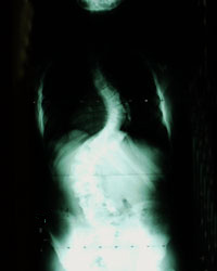
What Does An X-Ray Look Like?
An x-ray is a picture created in many shades of black, gray and white. Bones and other calcified structures appear almost white because they are denser than soft tissue and absorb more of the electromagnetic radiation. Muscle, fat and other soft tissues appear darker (black to gray tones) on the x-ray.