PLIF, ALIF, TLIF and XLIF:
Alphabet soup or surgical treatments for the spine?
Spinal fusion is a surgical procedure in which two or more vertebrae are joined or fused together. Fusion surgeries typically require the use of bone graft to facilitate fusion. This involves taking small amounts of bone from the patient’s pelvic bone (autograft), or from a donor (allograft), and then packing it between the vertebrae in order to "fuse" them together. This can be accomplished either posteriorly or between the vertebral bodies. When it is done between vertebral bodies, bone graft, along with a biomechanical spacer implant, will take the place of the intervertebral disc, which is entirely removed in the process. Spinal fusion surgery is a common treatment for such spinal disorders as spondylolisthesis, scoliosis, severe disc degeneration, or spinal fractures. Fusion surgery is usually considered only after extensive non-operative therapies have failed. Common fusion surgeries available include posterior fusion and interbody fusion such as PLIF, ALIF, TLIF or XLIF.
PLIF
PLIF stands for Posterior Lumbar Interbody Fusion. In this fusion technique, the vertebrae are reached through an incision in the patient’s back (posterior). The PLIF procedure involves three basic steps:
- Pre-operative planning and templating.
Before the surgery, the surgeon uses MRI and CAT scans to determine what size implant(s) the patient needs. - Preparing the disc space.
Depending on the number of levels to be fused, a 3-6 inch incision is made in the patient’s back and the spinal muscles are retracted (or separated) to allow access to the vertebral disc. The surgeon then carefully removes the lamina (laminectomy) to be able to see and access the nerve roots. The facet joints, which lie directly over the nerve roots, may be trimmed to allow more room for the nerve roots. The surgeon then removes the affected disc and surrounding tissue and prepares bone surfaces of adjacent vertebrae for fusion. - Implants inserted
Once the disc space is prepared, bone graft, allograft, or BMP with a cage (a biomechanical spacer implant) is inserted into the disc space to promote fusion between the vertebrae. The implant (cage) may be made of bone, metal, carbon fiber or other material. Additional instrumentation (such as rods or screws) will also be used at this time to further stabilize the spine. We do not recommend this procedure without using fixation because of high complications.
TLIF
TLIF stands for Transforaminal Lumbar Interbody Fusion. This fusion surgery is a refinement of the PLIF procedure and has recently gained popularity as another technique of surgical treatment for conditions affecting the lumbar spine. The TLIF technique involves approaching the spine in a similar manner as the PLIF approach but more from the side of the spinal canal through a midline incision in the patient’s back. This approach reduces the amount of surgical muscle dissection and minimizes the nerve manipulation required to access the vertebrae, discs and nerves. The TLIF approach is generally less traumatic to the spine, is safer for the nerves, and allows for minimal access and endoscopic techniques to be used for spinal fusion.
As with PLIF and ALIF, disc material is removed from the spine and replaced with bone graft (along with cages, screws, or rods if necessary) inserted into the disc space. The instrumentation helps facilitate fusion while adding strength and stability to the spine. We currently use many state of the art cage technologies including those made of bone, titanium, polymer, and even bioresorbable materials.
ALIF
ALIF stands for Anterior Lumbar Interbody Fusion. This procedure is similar to PLIF, however it is done from the front (anterior) of the body, usually through an incision in the lower abdominal area or on the side. This incision may involve cutting through, and later repairing, the muscles in the lower abdomen. At our practice, a mini open ALIF approach is available that preserves the muscles and allows access to the front of the spine through a very small incision. This approach maintains abdominal muscle strength and function and is oftentimes used to fuse the L5-S1 disc space.
Once the incision is made and the vertebrae are accessed, and after the abdominal muscles and blood vessels have been retracted, the disc material is removed. The surgeon then inserts bone graft (and anterior interbody cages, rods, or screws if necessary) to stabilized the spine and facilitate fusion.
XLIF
XLIF stands for eXtreme Lateral Interbody Fusion. This is a relatively new minimally invasive approach to the anterior spine that avoids an incision that traverses the abdomen and also avoids cutting or disrupting the muscles of the back. In this fusion technique, the disk space is accessed from a very small incision on the patient’s side (flank) a couple of inches in length, occasionally with another small incision (one inch long) just behind the first incision. Special retractors are utilized, in addition to a fluoroscopy machine, which provides real-time x-ray images of the spine. In addition, special monitoring equipment is used to determine the proximity of the working instruments to the nerves of the spine. The disk material is removed from the spine and replaced with a bone graft, along with structural support from a cage made of bone, titanium, carbon-fiber, or a polymer. This technique typically allows a shorter hospital stay and may be less painful than traditional approaches to the spine, however it also has limitations. Only those vertebra of the spine that have clear access from the side of the body can be approached using this technique. Also, only one or two levels can usually be accessed via this method.

Watch Animation about this Procedure
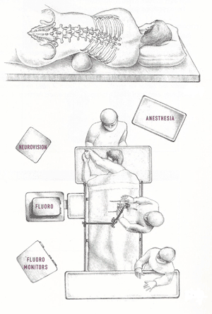
XLIF - patient positioning for surgery.
Fig 1: Above, Patient positioning during XLIF procedure.
Operating room layout for XLIF procedure.
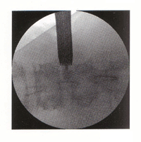
Fig 3: (above) Actual fluoroscope x-ray image
of retractor in place (posterior view).
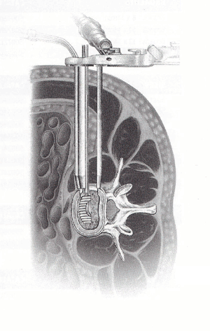
XLIF - Minimally Invasive Procedure Image.
Fig 4: XLIF retractor opened with bone graft
cage being placed in disk space (superior view).
(All above figures courtesy of Nuvasive, Inc., San Diego, CA)
Minimal Access
There are several types of spinal procedures utilizing minimal access techniques. The development of these techniques originated with the application of endoscopy during microdiscectomy surgery for herniated lumbar discs. It has now been applied to fusion surgeries. Ask a member of our clinical team to see if this might be right for you.
After Fusion Surgery
Recovery time is different for every patient. However, most patients are up and walking by the end of the first day after surgery. Most patients can expect to stay in the hospital for 3-5 days depending on their condition. Once released from the hospital, patients who have undergone surgery are given a prescription for pain medications to be taken if needed, as well as a detailed post-operative activity, physical therapy/exercise plan to help ease recovery and return to a healthy life.
